WE ARE CLOSING FOR AN UPGRADE!
The MNACTEC will be closed for improvement work until 17 September 2026.
We will still be busy with activities for schools,, online resources and on social media!
Ultrasound: from nature to medicine
The exhibition shows what ultrasound is, and how it is used in diagnostic tests and medical treatments.
From September 18, 2026
This travelling exhibition aims to explain what ultrasound is, and also how it is used for diagnosis and treatment in medicine.
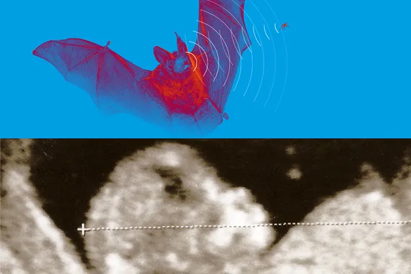
“Ultrasound: from nature to medicine" illustrates how observations of the natural world, maritime navigation, the study of the seabed and industrial needs have facilitated the development of ultrasound in the study of the human body, and its application in innovative medical treatments.
Ultrasound is these days used in medicine for such diagnostic tests as imaging scans, contrast-enhanced imaging and elastography, and in therapeutic treatments such as physiotherapy and rehabilitation, lithotripsy (breaking down the hardened masses known as kidney stones), while high-intensity focused ultrasound is soon expected to be available to break down tissues and tumours.
The exhibition includes a range of activities allowing the public to experiment with ultrasound. The most striking feature is the reproduction of a bat cave, which visitors enter with their eyes completely blindfolded, and have to find their way to the exit using an ultrasound device which simulates the echolocation used by bats.
The exhibits are divided into four areas:
1. Observation of nature
2. Physical principles
3. Diagnostic applications
4. Therapeutic applications
“Ultrasound: from nature to medicine" is an exhibition produced by the Radiology Service of Vall d'Hebron through the FUSMED research facility, which studies the therapeutic effects of high-intensity focused ultrasound, and forms part of the Molecular Medical Imaging unit of the Vall d'Hebron Institute of Research (VHIR). The exhibition was devised by Dr Xavier Serres, a radiodiagnostic imaging physician at Vall d’Hebron and researcher at the VHIR Molecular Medical Imaging research group.